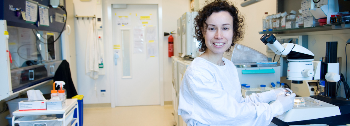
Around forty microscopes, several culture laboratories, and some 15,000 zebrafish: that is the workplace of Sylvia le Dévédec. She is one of the managers of the Leiden Cell Observatory, a unique facility that is open to all researchers.
Le Dévédec walks enthusiastically down the long corridor, pointing out various labs and rooms filled with microscopes. At the end is the zebrafish facility. Along the way, she repeatedly runs into students and colleagues and stops for a chat. “You meet people here from all kinds of disciplines, and that is what makes this work so valuable.”
Together with biologist Joost Willemse and chemist Amit Cherian, the LACDR researcher manages the microscopy section of the Cell Observatory: a shared facility where anyone who needs a microscope can come. As Le Dévédec explains, it makes little sense for different departments to buy the same expensive microscope when it can be shared instead. “That way, we create a diverse range of equipment that allows us to answer more research questions.”
“Life is incredibly beautiful”
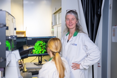
In the Cell Observatory’s annual photo competition, researchers compete for the title of most beautiful image. “I’ve had many wow moments behind the microscope myself,” says Le Dévédec. “You can see living cells moving with your own eyes, it’s incredibly beautiful.”
Metastatic cancer cells under the microscope
Le Dévédec herself researches breast cancer, with the ultimate aim of slowing down metastasis. How do cancer cells spread throughout the body? That is where microscopy comes in. “I try to understand the role of certain genes in cell migration,” she explains. “In a 96-well plate; a plastic plate about the size of a hand, with 96 small wells, we can test a different condition in each well. If you see the phenotype, in other words, the visible characteristics, change under the microscope, then you know that gene has played a role.”
A huge leap in innovation in just one generation
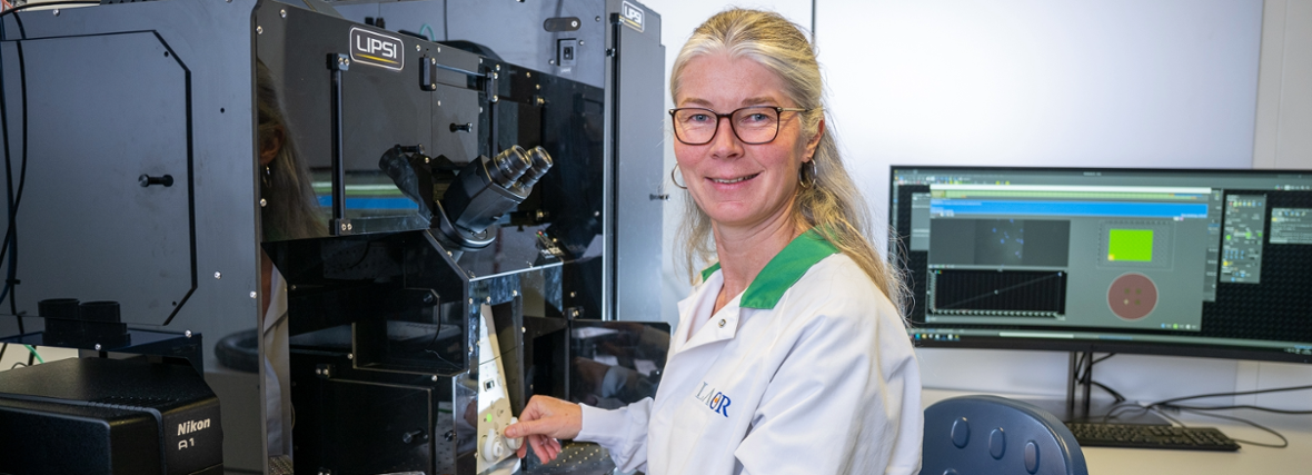
Things were very different during her PhD, Le Dévédec explains. “Back then, I could only study one condition at a time, because I had to manually track a few cells under the microscope all day. Now, in a single weekend, we can automatically photograph up to a million cells.”
She points to a microscope stacked with an entire tower of 96-well plates. These are kept in a space with the right temperature and humidity to keep the cells alive. A robotic arm continuously picks up the next plate and places it under the microscope, which then moves across each well and takes images. As Le Dévédec puts it: “There is so much possible nowadays.”
Microscopy brings science to life
As manager of the facility, she is also deeply involved in supporting its users — from advising on which microscope to use to helping secure funding for new equipment. She would also like to collaborate more with companies at Leiden Bio Science Park: “For small companies, it is often too expensive to purchase this kind of microscopy equipment themselves. I would love to offer them the opportunity to use our facilities and benefit from our experience.”
Whether she is talking about her own research, supervising PhD candidates, or managing the Cell Observatory, her passion is unmistakable. “Microscopy brings science to life,” says Le Dévédec. “It’s not just numbers, you can actually see what is happening in living cells. That is why I go to work with pleasure every day.”